Typical Animal Cell Diagram Labeled / Plant Vs Animal Cells Venn Diagram Labeled Diagram Of Plant Cell And Animal Cell Transparent Png 1675x1525 Free Download On Nicepng / It is easier to describe these parts by using diagrams:
byGertrud Landt-
0
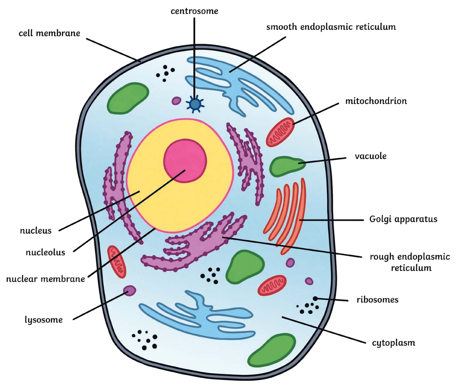
Typical Animal Cell Diagram Labeled / Plant Vs Animal Cells Venn Diagram Labeled Diagram Of Plant Cell And Animal Cell Transparent Png 1675x1525 Free Download On Nicepng / It is easier to describe these parts by using diagrams:. Draw a neat labelled diagram of animal cell. It is easier to describe these parts by using diagrams: Diagram of a typical animal cell, with the important features labeled. Bundle of microtubules to help separate the chromosomes during cell division. 5th grade science and biology.
Easy to navigate and repeat as needed until i felt confident in retaining the information! Featured in this printable worksheet are the diagrams of the plant and animal cells with parts labeled vividly. Animal cells come in all kinds of shapes and sizes, with their size ranging from a few millimeters to micrometers. Under the microscope, an animal cell shows many different parts called organelles, that work together to keep the cell functional. In this video the parts of an animal cell and its organelles are described.
What Is An Animal Cell Definition And Functions Twinkl from images.twinkl.co.uk Upvote (0) was this answer helpful? For more anatomy content please follow us and visit our website: Under the microscope, an animal cell shows many different parts called organelles, that work together to keep the cell functional. Illustration of the process by which somatic cells multiply and divide. For more anatomy content please follow us and visit our website: Mitosis is a process of cell division which results in the production of two daughter cells from a single parent cell. Diagram of a typical animal cell, with the important features labeled. Animal cells have a basic structure.
5th grade science and biology.
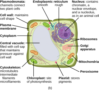
Typical plant and animal cells diagram 1 8 diagram quizlet image information: Though this animal cell diagram is not representative of any one particular type of cell, it provides insight into the primary organelles and the intricate internal structure of most animal cells. Protective and provide shape and size. Illustration of the process by which somatic cells multiply and divide. Found only in plant cells. Plant cell diagram | animal cell diagram. As observed in the labeled animal cell diagram, the cell membrane forms the confining factor of the cell, that is it envelopes the cell constituents together and gives the cell its shape, form, and existence. Bundle of microtubules to help separate the chromosomes during cell division. Diagram of a typical animal cell, with the important features labeled. Animal cells have a basic structure. For more anatomy content please follow us and visit our website: Easy to navigate and repeat as needed until i felt confident in retaining the information! 5th grade science and biology.
The typical animal cell format created a fun environment to learn the material. Bundle of microtubules to help separate the chromosomes during cell division. An animal cell ranges in size from 10 to 30 µm. For more anatomy content please follow us and visit our website: Labelled diagram of a typical animal cell animal4 {label gallery} get some ideas to make labels for bottles, jars, packages, products, boxes or classroom activities for free.
3 3 Eukaryotic Cells Concepts Of Biology 1st Canadian Edition from opentextbc.ca Humans are multicellular organisms with various different types of cells that work together to sustain life. 5th grade science and biology. Bundle of microtubules to help separate the chromosomes during cell division. Anatomynote.com found typical animal cell and plant cell diagram from plenty of anatomical pictures on the internet. Animal cell and plant cell from 3.bp.blogspot.com this activity is a simple reinforcement worksheet to help students learn the structures found within a typical animal cell and what those structure might look like on a this can also be assigned with the. Animal cell diagram (label) study. Below the basic structure is shown in the same animal cell, on the left viewed with the light. Typical plant and animal cells diagram 1 8 diagram quizlet image information:
Cells are made up of different parts.
Fungal cell wall is made up of chitin (not cellulose). Cells are one of the most basic building blocks of life. Featured in this printable worksheet are the diagrams of the plant and animal cells with parts labeled vividly. Plant cell diagram | animal cell diagram. In a typical animal cell, mitosis can be divided into four principals stages: Click here👆to get an answer to your question ️ draw a neat labelled diagram of animal cell. The daughter cells are identical to one another and to the original parent cell. Found only in plant cells. Large, hairlike structure used for the movement of some cells. Easy to navigate and repeat as needed until i felt confident in retaining the information! In this video the parts of an animal cell and its organelles are described. Illustration of the process by which somatic cells multiply and divide. The typical animal cell format created a fun environment to learn the material.
We think this is the most useful anatomy. An easy and convenient way to make label is to generate some ideas first. Cells are one of the most basic building blocks of life. For more anatomy content please follow us and visit our website: Below the basic structure is shown in the same animal cell, on the left viewed with the light.
Plant Cell Diagram By Russell Kightley Media from rkm.com.au As observed in the labeled animal cell diagram, the cell membrane forms the confining factor of the cell, that is it envelopes the cell constituents together and gives the cell its shape, form, and existence. The typical animal cell format created a fun environment to learn the material. Diagram of a typical animal cell, with the important features labeled. Click here👆to get an answer to your question ️ draw a neat labelled diagram of animal cell. Diagram of plant and animal cell. Below the basic structure is shown in the same animal cell, on the left viewed with the light. Animal cell and plant cell from 3.bp.blogspot.com this activity is a simple reinforcement worksheet to help students learn the structures found within a typical animal cell and what those structure might look like on a this can also be assigned with the. Draw a neat labelled diagram of animal cell.
Easy to navigate and repeat as needed until i felt confident in retaining the information!
Page 3 a typical animal cell 1. Posted by donna steinke on 6/9/2009 12:00:00 am reply The typical animal cell format created a fun environment to learn the material. Introduction and terminology | basic human anatomy / 598 x 530 pixel type jpg download.smallest living structure and constituent unit of all animals there should be no lasting side effects of the miniaturization, except, i hope, a slight tingling sensation caused by new knowledge and a growing excitement about what scientists know—and still don. Illustration of eukaryotic and prokaryotic cell with text. However, the cell membrane in plant cells is quite rigid, while, the cell membrane in animal cells is quite flexible. Protective and provide shape and size. We hope this picture structure of a typical animal cell diagram can help you study and research. Related for labelled diagram of a typical animal cell labeled diagram of arm veins | diagram labels {label gallery} get some ideas to make labels for bottles, jars, packages, products, boxes or classroom activities for free. Below the basic structure is shown in the same animal cell, on the left viewed with the light. Bundle of microtubules to help separate the chromosomes during cell division. Almost all animals and plants are made up of cells. Animal cell anatomy diagram structure with all parts nucleus smooth rough endoplasmic reticulum cytoplasm golgi apparatus mitochondria membrane centro prokaryote vs eukaryote.
We think this is the most useful anatomy typical animal cell diagram. Cells are one of the most basic building blocks of life.